2. Brain Beginnings - Update 03 Septr 2021
The following column shows the content of the chapter in order as it appears. Use search & find or scroll down to locate what you want.
Embryology of the Nervous System; Its Origin
Development of Brain
Orienting the Anatomic Terms
Embryology of the Nervous System; Its Origin: Imagine looking down on an earthworm’s long tubular fleshy back. The early embryo of humans, if straightened, would resemble it. The formation of a section of the neural tube from the original neural plate along the back of the early embryo is shown in the below figure.
Neural tissue represents a branching off of a segment of skin surface (ectoderm) tissue which forms a neural plate along the back of the early embryo.

Neural tissue represents a branching off of a segment of skin surface (ectoderm) tissue which forms a neural plate along the back of the early embryo.
The neural plate folds to form the neural tube.
(Electron micrographs of section of chick neural tube.)
A.
Early-on, 3 germ cell layers—the outer surface ectoderm, the middle-layer mesoderm,
and the inner-surface endoderm—lie close together. The ectoderm neural
plate is precursor of the central and peripheral nervous systems.
B. The neural plate buckles at its mid line.
C. Closure of the dorsal neural folds forms the neural tube.
D.
The neural tube lies over the notochord and is flanked by somites, various level
ovoid groups of mesodermal cells that give rise to muscle and cartilage.

As the above shows, at 1 week after conception, the skin of the embryo's back forms a ridge like a surface canal, then a top open tube, and then the open tube sinks in and closes to form the neural tube that extends just under the back of the early embryo from its head part. The cavity and cells that make the tube's wall and lining are the forerunners of the brain (the front part) and spinal cord (the tail). In adult development, the upper tube becomes the brain ventricles and the lower part is spinal cord and retains the cavity as a tiny central canal.
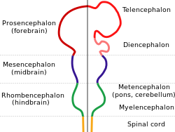
The neural tube nearest the head of the embryo, which will become the brain, by cell multiplication, enlarges like an expanding bubble to form the early brain vesicles you see here. These developments are the result of numerous neural development stimulators, chemicals that direct the pathways and shapes nd locatiuns of neurons and glia in the nervous system. They can be easily disturbed by external influences.
 Development of Brain: The part of the prosencephalon that becomes left and right cerebral gray-matter, cerebral-cortex-covered hemispheres and white-matter subcortex of the brain is called telencephalon. Each side contains hollow-tube, fluid-filled left and right ventricle that connects. The cerebral cortex is the functional body of conscious mind. Consciousness is not yet formed at birth; the interaction of the body with its environment results in a full consciousness by several years after birth and it continues to develop throughout life. It is a function of the whole nervous system interacting. It is not an anatomical body.
 Most of the cerebral cortex develops in the adult into a 6-layer, millimeters-thick cover of connected vertical modules, each module formed by millions of nerve cells (neurons) that migrate to the brain's surface from parent cells that lined the brain's primitive ventricles. The cerebral cortex might be imagined as the thin red strip in above figure that outlines the telencephalon; it contains the gray matter because its cells make it darker than the underlying much larger nerve fiber inner core, which is the white matter. The brain ventricles secrete cerebrospinal fluid (CSF). There is little or no entry from the blood into the CSF and even when the blood vessels are up against the ventricle wall, a blood-brain barrier prevents the exchange of particles. The white matter within the telencephalon is bundles of nerve fibers connecting the cerebral cortex neurons (nerve cells) between themselves and to the subcortical and lower level (lower brain and spinal cord) neurons.
Behind and between the telencephalon is the "bridging brain," or diencephalon, which develops into right and left thalamus and the underlying hypothalamus, and has the pineal gland. The thalamus is the brain's central switching center, transmitting signals between the cerebral cortex and subcortical bodies like the basal ganglia and the separate cerebellum, and it also connects incoming signals from the peripheral sensory nerve structures and distributes these signals to the cerebral cortex.
Behind and between the telencephalon is the "bridging brain," or diencephalon, which develops into right and left thalamus and the underlying hypothalamus, and has the pineal gland. The thalamus is the brain's central switching center, transmitting signals between the cerebral cortex and subcortical bodies like the basal ganglia and the separate cerebellum, and it also connects incoming signals from the peripheral sensory nerve structures and distributes these signals to the cerebral cortex.
Latest research shows that recurrent circuits between sections of thalamus and cerebral cortex at around 40 hz wave rate modulate our consciousness. It means that consciousness is located by a electromagnetic rhythm and not by a place.
The diencephalon's pineal gland secretes the sleep hormone melatonin into the cerebrospinal fluid at night or in absence of light and the diencephalon's hypothalamus influences emotions, eating, drinking, sleeping and all the endocrine glands. The pituitary gland is a downward extension of the hypothalamus and is the master gland of all the other body glands by the influence it exerts on them by stimulating all the other body hormones.
The diencephalon and midbrain also contains many of the brain's other subcortical structures and most of the the basal ganglia. and smaller nuclei of neurons. And keep in mind that the diencephalon structures each have a left and right except the pineal gland and the hypothalamus, each of which is a single mid line bilaterally symmetrical structure. The diencephalon surrounds the brain’s 3rd ventricle, and the 1st and 2nd ventricles are at the cores of the left and right cerebral hemispheres that pass the cerebrospinal fluid from the cerebral ventricles down towards the 4th ventricle and into the spinal cord central canal.
The mesencephalon becomes the midbrain (section of Brain in between lower border of Thalamus and upper border of Pons), which gets packed with nerve fibers carrying signals to and from the upper brain to lower brain and spinal cord. It contains many small nuclei of neurons and the upper cranial nerves. It is much involved in eye movements and control of vital organ functions like respiration, heart beat, digestion and excretion.
The rhombencephalon, or hindbrain surrounds the 4th ventricle and its front part grows to become the pons ("Bridge" in Latin; so called because it is made up almost completely of laterally crossing fibers that form a bridge between left and right side) and, its rear part grows to become the cerebellum (perched atop and on the back of the pons and atop the 4th ventricle between the back of the pons and the front of the cerebellum). The connections between cerebrum and lower brain develop as thick bundles of connecting fibers in the adult - the peduncles.
And the lowest part, the myelencephalon, becomes brain's medulla and connects the brain above with the spinal cord below. The medulla is the important site of the neuron nuclei that control breathing, modify heart action and blood pressure, and have strong effects on the autonomic nervous system. The aforementioned cranial nerves have their lower nuclei in the upper medulla.
The Anatomy - Planes and other Terms:
The Planes: Descriptions and figures may refer to slices of brain or other anatomy. For the usual reader, refer to the 3-dimensions - the horizontals width and length, and the verticals up-down.
The transverse (aka "axial") is any horizontal slice, horizontal front-back and left-right.
The sagittal, is a vertical front-back slice. Sagittal alone means down the center of the body from front to rear while parasagittal are parallel plane slices to left or right of the sagittal.
The coronal is a vertical slice at right angle to the sagittal. It may be visualized as left to right, ear to ear slice with the outline of a king's crown atop a head, face forward.
The terms rostral, caudal, midbrain, brain stem, pons, medulla (medulla oblongata) are better located by the following.
Rostral (higher up) and Caudal (toward the rear or the tail) give the orienting direction from up-down or higher to lower. In the human brain, the cerebral cortex, which is part of the left and right cerebral hemispheres or cerebrum, is most rostral. Then, in a successively caudal direction comes the diencephalon, which contains the right and left thalamus and, beneath it, the hypothalamus. Then the midbrain, which contains structures (3rd ventricle, the more caudal basal ganglia like substantia nigra, etc.) between the diencephalon and the next caudal structure, the pons, in front and the cerebellum, which sits atop the rear of the pons.
Then, caudal to the pons is the medulla oblongata ("the medulla") that connects brain with spinal cord.
The term, brain stem, includes, from rostral to caudal, the midbrain, the pons, and the medulla (these 3 structures being in a direct line connecting the upper brain - cerebrum and diencephalon - rostrally with the spinal cord, caudally.
Finally, readers should keep in mind that the development of the nervous system described above hinges on a hugely cooperative control system described as follows:
Early
in development ectodermal cells are firstly faced with the choice of whether to
become neural or epidermal (skin) cells. This decision is the most
fundamental step of neural development. Much research has
focused on a search for signals that control this development.
We
now know that two major classes of proteins work together to promote
the differentiation of an ectodermal cell into a neural cell. The first
are inductive factors,
signaling molecules that are secreted by nearby cells. Some of these
factors are freely diffusible and exert their actions at a distance, but
others are tethered to the cell surface and act locally. The second inductive factor is
surface receptors that enable cells to respond to inductive factors.
Activation of these receptors triggers the expression of genes that
encode intracellular proteins—transcription factors, enzymes, and
cytoskeletal proteins—which push ectodermal cells along the pathway to
becoming neural cells.
The ability of a cell to respond to inductive signals, termed its competence,
depends on the repertory of receptors, transduction molecules,
and transcription factors expressed by the recipient cell. Thus a cell's
fate is determined not only by the signals to which it is exposed—a
consequence of when and where it finds itself in the embryo—but also by
the profile of genes it expresses as a consequence of its prior
developmental history.
What all of us should bring away from this is that this complex system may be disturbed by many factors affecting the pregnant woman: x-ray, an MRI's magnetic pulls, vitamins (lack or or too much) toxins (alcohol, tobacco and coffee). Thus, the importance of shielding pregnancy from outside effects and maintaining the pregnant woman in a virtual cocoon. Also very important to keep in mind are that the potentially bad affects are analog rather than digital. It means: rather than an all or none destructive effect, these bad effects are more or less and this probably explains why individuals differ from other individuals in many good and bad qualities like intelligence or sports ability or musical talents.
We know development down to a certain submicroscopic point, but below that point, it is still a mystery. I do not know about you, but I love a mystery!
End of Chapter. To read next, click 9.3 Secrets of the Brain tissues - A Starter
2 comments:
I think this is among the most vital information for me and I am satisfied reading your article.
https://blog.mindvalley.com/midbrain-function
Found your post interesting to read. I can’t wait to see your post soon. Good Luck for the upcoming update. This blog is really very interesting and effective.
https://blog.mindvalley.com/diencephalon/
Post a Comment